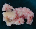
This procedure is indicated in patients with prolapse intervertebral disc not responding to non-operative treatment or with progressive neurological deficit. This procedure is performed under general or regional anesthesia. The location of disc is confirmed pre-operatively with X-rays and MRI Scan.
The operation is performed with the patient’s face lying down (prone position). A small incision is taken at midline corresponding to the level of prolapsed disc. The paraspinal muscles are reflected and the level of disc prolapse is confirmed with help of a special x-ray machine called image intensifier. A microscope and specialized retractors are used to aid illumination and magnification. The ligament between the two laminae called as ligamentum flavum or yellow ligament is incised. The nerve root, which is compressed is identified and retracted. The proplased disc is then removed after incising the outer covering, the annulus fibrosus. In some patients the disc may have protruded out of the annulus and may be lying free in the canal. The nerve is freed which is the main intention of the procedure. The incision is then closed in layers.
Patient can start mobilizing same day following surgery. The success rate for this procedure for relieving leg pain is 90%. The complications following this procedure are infection and damage to nerve root or the covering of nerves called dura mater. The incidence of these complications is low but patient needs to be aware of the possible risks before undergoing this procedure.